
Scientific Computing and Data / Research Data Services / Data Ark: Data Commons / Digital Pathology Slides
AIR·MS: De-identified Digital Pathology Slides
Overview
The Department of Pathology, Molecular and Cell-Based Medicine, and Scientific Computing and Data are pleased to announce the incorporation of linked data into our research repository of over 2.5 million de-identified digital pathology slides. This new data linking, which connects de-identified patient information with digital images of pathology slides (stored on the Minerva supercomputer) will make it possible to perform image analysis and use machine-learning or artificial intelligence at scale across millions of slides, and open exciting opportunities for research and discovery. The linked slides are accessible to any user who agrees to a Data Use Agreement and can be accessed through AI Ready Mount Sinai (AIR·MS).
This extensive collection represents a wide range of organ systems, such as lung, heart, pancreas, kidney, liver, gastrointestinal, genitourinary, gynecological, hematological, and neuropathological systems. The repository encompasses a broad spectrum of disease processes, such as neoplastic, developmental, inflammatory, toxic, metabolic, genetic, degenerative, traumatic, and infectious pathologies.
Slides have been prepared using diverse staining techniques, ranging from routine hematoxylin and eosin (H&E) to specialized stains such as silver, trichrome, and immunohistochemistry. Together, this rich and diverse data set provides a powerful resource for advancing the study of human disease through digital pathology.
To learn more about Digital Pathology Slides and how to use them in your research, be sure to join the upcoming training session on November 12, 2026, 10.00-11.00 a.m. EST!
Register in advance for this webinar: https://tinyurl.com/yc5v3d57
After registering, you will receive a confirmation email containing information about joining the webinar.
What Is Hosted Under AIR·MS?
Currently, AIR·MS hosts over 2.6 million de-identified digital pathology slides linked with de-identified patient information, with more becoming available on a continuing basis. The slides were scanned on the Philips Ultrafast at 40x magnification. iSyntax files were converted to TIF. Slides served through AIR·MS have been de-identified and digitized into a readable TIF format.
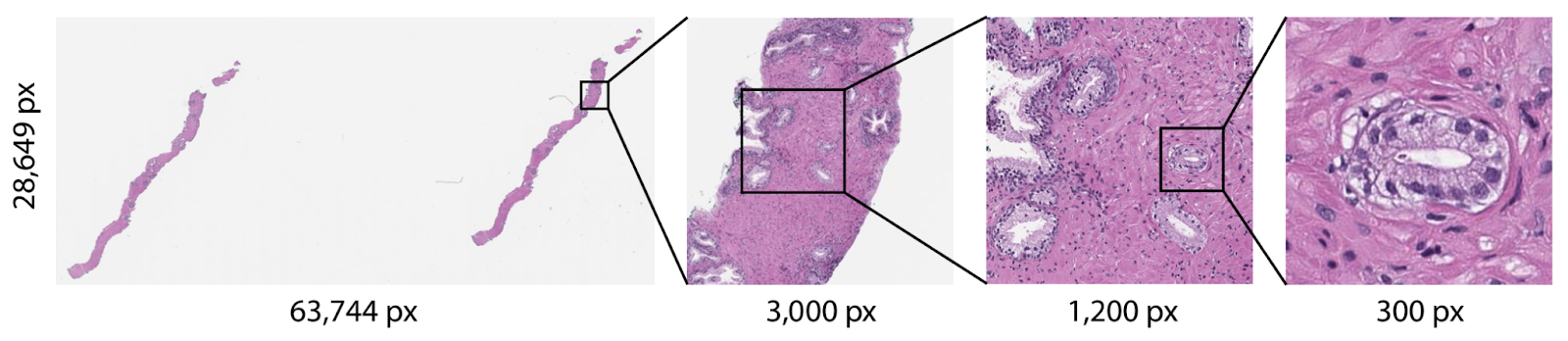
Figure 1. Digitized pathology slides are gigapixel images that can span hundreds of thousands of pixels in each dimension.
Example: Digital pathology slides collected from 191,119 patients through October 1, 2024, with demographic information detailed below.
Table 1. Cohort demographics: (a) gender, (b) race and ethnicity, and (c) age.
| Gender | Count |
|---|---|
| Female | 120,121 |
| Indeterminate | 13 |
| Male | 70,945 |
| Unknown | 40 |
| Race Ethnicity Combined | Count |
|---|---|
| American Indian or Alaska Native | 140 |
| Asian | 14,071 |
| Black or African-American | 28,323 |
| Hispanic | 35,508 |
| Native Hawaiian or Pacific Islander | 159 |
| Patient Declined | 958 |
| White | 66,515 |
| Unknown/Other/Not Reported | 45,445 |
| Age Group | Count |
|---|---|
| 0-10 | 1,377 |
| 11-20 | 3,239 |
| 21-30 | 14,950 |
| 31-40 | 28,684 |
| 41-50 | 31,704 |
| 51-60 | 35,551 |
| 61-70 | 38,128 |
| 71-80 | 26,597 |
| 81-90 | 9,424 |
| 91+ | 1,465 |
The digital pathology technology effectively reduces process time
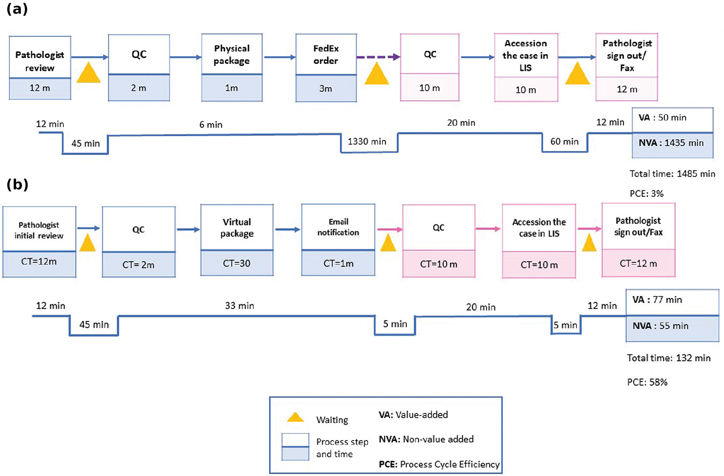
Figure 1. Value stream mapping of simple (a) traditional glass slide vs. (b) digital slide consult. Image adapted from Haghighi, Mehrvash, et al. 2021. doi: 10.4103/jpi.jpi_74_21.
Access
- To use this data, you must read, agree and sign the Data Use Agreement (you must be logged in through the Mount Sinai campus network or secure remote VPN). IRB (Institutional Review Board) approval is not required to access Digital Pathology Slide data via AIR·MS.
- After access has been granted, you can view slides by going to the folder on Minerva directly. To get the path variable, you can load the module by issuing the command $ module load dataark.
- Further details about how to use these slides in your research are available here with an accompanying video recording here.
- If you have questions, you can submit a ticket here: https://hpims.atlassian.net/servicedesk/customer/portal/67
Further Information
- To access de-identified and protected health information (PHI) versions of the digital pathology slides using Python follow these links:
- For more information on the Department of Pathology and Laboratory Medicine, please visit https://www.mountsinai.org/patient-care/service-areas/pathology.
- For more information on the Minerva high-performance computing and data ecosystem including the Data Ark data commons, please visit https://labs.icahn.mssm.edu/minervalab/ and https://labs.icahn.mssm.edu/minervalab/resources/data-ark/.