
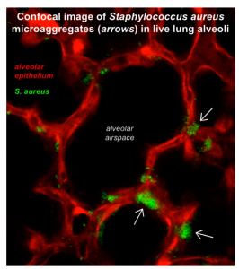
PROJECT 1: Staphylococcus aureus, a major lung pathogen, rapidly forms microaggregates in structural niches of lung alveoli to stabilize, initiate alveolar infection, induce alveolar damage, and resist antibiotic therapy.
S. aureus bacteria cause severe pneumonia in previously healthy people, but how the inhaled bacteria stabilize in alveoli – the lung’s air sacs – to initiate a lung infection is not clear. Our lab discovered that contact with the alveolar epithelium causes inhaled S. aureus (green fluorescence in the confocal microscopic image above) to form microaggregates (arrows) in structural, corner-like niches of alveolar walls (red fluorescence). Microaggregate formation allows the bacteria to stabilize in the protected niche locations, where they secrete bacterial toxins that damage the epithelium. The damage signals then spread through epithelial gap junctions to neighboring, uninfected alveoli, leading to fluid leakage into alveolar airspaces, respiratory failure, and death in mouse models.
We developed two new therapeutic approaches that protect against S. aureus-induced lung injury in mice: (1) disrupting the PhnD protein interactions that stabilize microaggregated S. aureus; and (2) blocking the epithelial gap junctions that spread microaggregate-induced alveolar damage signals. You can find the paper here and a video version of the abstract here.
The novelty of these findings center on a new role for alveolar microanatomy in lung disease pathogenesis and drug efficacy. Importantly, this project challenges the notion that the extensive spatial profile of bacteria-induced lung injury is caused by widespread lung infection or immune activation. Rather, lung injury may result from bacterial toxin-induced damage to niche-localized alveolar walls, which is amplified to healthy alveoli by cell-to-cell spread of the damage signals. We think this concept might explain the clinical observation that localized S. aureus lung infection can quickly progress to severe pneumonia despite antibiotic therapy.
PROJECT 2: Influenza promotes secondary pneumonia by Staphylococcus aureus by disrupting alveolar liquid secretion, an important alveolar defense.
Death among people with influenza infection is often caused by a secondary pneumonia by inhaled S. aureus bacteria, but how influenza promotes S. aureus coinfection is not understood. Our lab discovered that influenza infection disrupts alveolar liquid secretion, which is a normal, CFTR-dependent epithelial function that usually prevents inhaled S. aureus from stabilizing against alveolar walls and promotes their clearance toward the airways. In the absence of alveolar liquid secretion, inhaled S. aureus can gain a foothold in alveoli to initiate coinfection and cause lung injury. However, rescue of alveolar liquid secretion by the CFTR modulator drug, ivacaftor, restored bacterial clearance and prevented fatal coinfection-induced lung injury in influenza-infected mice. This paper can be found here.
These findings represent the first understanding of alveolar responses to influenza in situ. In addition, they identify a new role for alveolar liquid secretion in antibacterial host defense and raise the possibility that CFTR modulator drugs could protect against fatal bacterial coinfection in people with influenza infection.
Follow-up work from our lab discovered that CFTR in alveolar type 1 cells – a cell type not considered to be relevant to lung liquid dynamics or the pathogenesis of CFTR-related diseases – contributes to alveolar liquid secretion and host defense (here). Additional, preprint data from our lab reveal that alveolar liquid dynamics depend on lung perfusion and its effects on ion channels in the alveolar epithelium (here). Related reviews (here and here) highlight the therapeutic potential of CFTR modulator drugs and other host-directed approaches for severe lung infection.
PROJECT 3: Future therapy for lung injury might leverage endogenous epithelial repair mechanisms to promote lung healing after pneumonia.
Although respiratory viruses and bacteria can cause severe pneumonia and lung injury, the lung can often recover from the infection by repairing itself. State-of-the-art medical care tries to support the lungs and the body while the repair processes initiate and proceed on their own. But what if we could accelerate lung repair? Could we get our patients off respiratory support faster and home faster? Could we stimulate or rescue repair in people in whom pneumonia seems overwhelming? We’re trying to start to answer these questions now in our laboratory. We discovered recently that the alveolar epithelium can restore the lung’s air-blood barrier function after S. aureus infection through a mechanism that centers on the Notch protein. Briefly, Notch protein cleavage leads to exposure of the Notch transmembrane domain that stimulates junctional protein reassembly and rescues air-blood barrier function in preclinical models (see preprint here). We’re working to translate our findings to human tissue, with the long-term goal of developing a first epithelium-directed treatment for pneumonia. A related review (here) discusses known lung repair mechanisms, thereby highlighting the novelty of our preprint findings.
ONGOING RESEARCH PROJECTS
Our other ongoing research builds on our prior work. Funded projects focus on the endogenous mechanisms by which S. aureus-damaged alveoli are repaired and alveolar responses to influenza that promote coinfection by S. aureus and cause immune-mediated lung injury. Since lung infection disproportionately affects the young and the old, aspects of our ongoing work aim to address how alveolar responses to inhaled bacteria and viruses change across the lifespan.
A complete list of our publications can be found here.