Research
Clinical disability in multiple sclerosis (MS) is driven by immune cell infiltration into the CNS parenchyma. Understanding the mechanisms by which CNS barrier cells regulate immune cell activation and entry during this infiltrative process may identify novel therapeutic strategies for MS and other CNS autoimmune diseases.
During CNS inflammation, immune cells traffic from the blood through a two-barrier structure termed the neurovascular unit. While many have focused on entry through the first barrier, a specialized endothelial wall known as the blood-brain barrier, less is known about how immune cells interact with the second barrier, a layer of endfoot processes extending from specialized cells called astrocytes, a barrier referred to as the glia limitans.
In MS lesions, immune cells circulate within the space between the first and second barriers, a compartment termed the perivascular space. Within this space, immune cells interact with the astrocyte endfeet and potentially receive signals that prime them for autoimmune attack. Our laboratory is interested in identifying and characterizing the signaling pathways between astrocytes and immune cells within the perivascular spaces. We are testing the hypothesis that this cross-talk regulates immune cell function prior to CNS entry and induces functional differentiation in both cell types which contributes to both the acute phase of CNS inflammation and more chronic processes of neurodegeneration.
Sam Horng
Icahn 10-20A
New York, NY 10029
(212) 659-1692
sam.horng@mssm.edu
Publications
2023
2022
Satyanarayan S, Safi N, Sortes T, Filomena S, Zhang Y, Klineova S, Fabian M, Horng S, Tankou S, Miller A, Krieger S, Lublin F, Sumowski J, Katz Sand, I. Differential antibody response to COVID-19 vaccines across immunomodulatory therapies for multiple sclerosis (2022). Mult Scler Relat Disord. 2022 Mar 12; 62:103737. Doi: 10.1016/j.msard.2022.103737.
Amatruda M, Chapouly C, Woo V, Safavi F, Zhang J, Dai D, Therattil A, Moon C, Gordon A, Parkos, Horng S. Astrocytic Junctional Adhesion Molecule-A regulates T cell entry past the glia limitans to promote CNS autoimmune attack (2022). Brain Comm. 4(2): fcac044. doi: 10.1093/braincomms/fcac044 Epub 2022 Feb 18.
2021
George I, Arrighi-Allisan A, Delman BN, Balchandani P, Horng S* and Feldman R.* *Co-senior author. A novel method to measure venular perivascular spaces in multiple sclerosis patients on 7 Tesla MRI. (2021) Am J Neuroradiol. Jun;42(6):1069-1072. doi: 10.3174/ajnr.A7144. Epub 2021 Apr 15.
Sumowski JF, Horng S, Brandstadter R, Krieger S, Leavitt VM, Katz-Sand I, Fabian M, Klineova S, Graney R, Riley CS, Lublin FD, Aaron AE, Varga A. Sleep disturbance and memory dysfunction in early multiple sclerosis. (2021) Ann Clin Transl Neurol.Jun;8(6):1172-1182. doi: 10.1002/acn3.51262. Epub 2021 May 5.
2020
Mora P, Hollier PL, Guimbal S, Abelanet A, Diop A, Cornuault L, Couffinhal T, Horng S, Gadeau AP, Renault MA, Chapouly C. Blood-brain barrier genetic disruption leads to protective barrier formation at the Glia Limitans. (2020) PLoS Biol. Nov 30;18(11):e3000946. doi: 10.1371/journal.pbio.3000946. eCollection 2020 Nov. PMID: 33253145
2019
Jordan S, Tung N, Casanova-Acebes M, Chang C, Cantoni C, Zhang D, Wirtz TH, Naik S, Rose SA, Brocker CN, Gainullina A, Hornburg D, Horng S, Maier BB, Cravedi P, LeRoith D, Gonzalez FJ, Meissner F, Ochando J, Rahman A, Chipuk JE, Artyomov MN, Frenette PS, Piccio L, Berres M, Gallagher EJ, Merad M. (2019) Dietary intake regulates the circulating inflammatory monocyte pool. Cell. Aug 22;178(5):1102-1114.e17. doi: 10.1016/j.cell.2019.07.050.
2018
Harel A, Horng S, Gustafson T, Ramieneni A, Farber RS, Fabian M. (2018) Successful treatment of progressive multifocal leukoencephalopathy with recombinant interleukin-7 and maraviroc in a patient with idiopathic CD4 lymphocytopenia. J Neurovirol. doi: 10.1007/s13365-018-0657-x.
2017
Horng S, Therattil A, Moyon S, Gordon A, Kim K, Argaw AT, Hara Y, Mariani JN, Sawai S, Flodby P, Crandall ED, Borok Z, Sofroniew MV, Chapouly C, and John GR. (2017) Astrocytic tight junctions control inflammatory CNS lesion pathogenesis. J Clin Invest 1;127(8):3136-3151.
Horng S and Fabian M. (2017) The Pathophysiology and Presentation of Multiple Sclerosis. Handbook of Relapsing Remitting Multiple Sclerosis. Aaron Miller (Eds.)
Horng S. (2017) Impairments in Moral Decision Making due to Neurodegenerative Disease: Ethical and Practical Considerations for Clinicians. Journal of Hospital Ethics 4(2):65-73.
2016
Laitman BM, Asp L, Mariani JN, Zhang J, Liu J, Sawai S, Chapouly C, Horng S, Kramer EG, Mitiku N, Loo H, Burlant N, Pedre X, Hara Y, Nudelman G, Zaslavsky E, Lee YM, Braun DA, Lu QR, Narla G, Raine CS, Friedman SL, Casaccia P, John GR. (2016) The Transcriptional Activator Krüppel-like Factor-6 Is Required for CNS Myelination. PLoS Biol. 14(5):e1002467.
2015 & Earlier
Chapouly C, Argaw AT, Horng S, Castro K, Zhang J, Asp L, Laitman BM, Mariani JN, Zaslavsky E, Nudelman G, John GR. (2015) Astrocytic ECGF1/TP and VEGF-A drive blood-brain barrier opening in inflammatory CNS lesions. Brain. 138(Pt 6): 1548-67.
Horng S and Miller FM. (2013) Ethics of Sham Surgery in Clinical Trials for Neurologic Disease. Handbook of Neuroethics. Jens Clausen and Neil Levy (Eds.)
Merlin S, Horng S, Marotte LR, Sur M, Sawatari A, Leamey CA. (2013) Deletion of Ten-m3 induces the formation of eye dominance domains in mouse visual cortex. Cereb Cortex. 2013 Apr;23(4):763-74.
Horng S, Kreiman G, Ellsworth C, Page D, Blank M, Millen K, Sur M. (2009) Differential Gene Expression in the Developing Lateral Geniculate Nucleus and Medial Geniculate Nucleus Reveals Novel Functional Roles for Zic4 and Foxp2 in Visual and Auditory Pathway Development. J Neurosci. 29(43): 13672-83.
Horng S and Sur M. (2009) Patterning and Plasticity of Maps in the Mammalian Visual Pathway. In The Cognitive Neurosciences IV, Ed Gazzaniga. MIT Press, 2009.
Lyckman AW, Horng S, Leamey CA, Tropea D, Watakabe A, Van Wart A, McCurry C, Yamamori T, Sur M. (2008) Gene expression patterns in visual cortex during the critical period: synaptic stabilization and reversal by visual deprivation. Proc Natl Acad Sci U S A. 105(27):9409-14.
Horng SH, Miller FG. (2007) Placebo-controlled procedural trials for neurological conditions. Neurotherapeutics. 4(3):531-6. Review.
Horng SH and Sur M. (2006) Visual activity and cortical rewiring: activity-dependent plasticity of cortical networks. In Prog in Brain Res 157, Ed (Moller), Elsevier B. V., pp 3-11.
Tropea D, Kreiman G, Lyckman A, Mukherjee S, Yu H, Horng S, Sur M. (2006) Gene expression changes and molecular pathways mediating activity-dependent plasticity in visual cortex. Nat Neurosci. 9(5):660-8.
Horng S, Grady C. (2003) Misunderstanding in clinical research: distinguishing therapeutic misconception, therapeutic misestimation, and therapeutic optimism. IRB. 25(1):11-6.
Horng S, Miller FG. (2003) Ethical framework for the use of sham procedures in clinical trials. Crit Care Med. 31(3 Suppl):S126-30.
Horng S, Emanuel EJ, Wilfond B, Rackoff J, Martz K, Grady C. (2002) Descriptions of benefits and risks in consent forms for phase 1 oncology trials. N Engl J Med. 347(26):2134-40.
Horng S, Miller FG. (2002) Is placebo surgery unethical? N Engl J Med. 347(2):137-9.
Tobias ML, Barnard C, O’Hagan R, Horng S, Rand M and Kelley DB. (2004) Vocal communication between male Xenopus laevis. Animal Behaviour. 67: 353-365.
Kelley DB, Tobias ML and Horng S. (2001) Producing and perceiving frogs songs; dissecting the neural bases for vocal behaviors in Xenopus laevis. In Anuran Communication, M. Ryan (Ed), Smithsonian Institution Press, pp. 156 – 166.
Awards
- K08 Mentored Clinical Scientist Research Career Development Award, National Institute of Neurological Diseases and Stroke, National Institutes of Health, 2018 to Present
- Career Transition Award jointly funded by the National Multiple Sclerosis Society and the Conrad N. Hilton Foundation, Icahn School of Medicine at Mount Sinai Hospital, 2017 to 2018
- Leon Levy Fellowship in Neuroscience, 2015 to 2017
- R25 Research in Residency Fellowship, National Institutes of Health and Mount Sinai Hospital, 2015-2016
- Arnold P. Gold Foundation’s Humanism and Excellence in Teaching Award, Department of Medical Education at the Icahn School of Medicine at Mount Sinai, 2012-2013
- Harvard Medical School Medical Scientist Training Program, 2009-2011
- F30 Predoctoral NRSA Fellowship, National Institute of Neurological Diseases and Stroke, 2008-2010
- Aspen Institute Health Forum, Predoctoral Fellow, 2007
- Pre-IRTA Award, National Institutes of Health, Sept 2000
- Summa Cum Laude, Columbia University, May 2000
- Honors in Department of Biological Sciences, May 2000
- Gold Kings Crown Award, Columbia University, May 2000
- Organizational Achievement Award, Columbia University, May 2000
- Phi Beta Kappa, elected December 1999
Projects
1. How do CNS barrier cells regulate immune cell function during autoimmune attack?
The CNS evolved a unique two barrier system through which immune cells must penetrate in a step-wise fashion in order to access the parenchyma in a wide range of neurologic diseases. In multiple sclerosis, immune cells circulate within the space between the first (vascular) and second (glial) barriers, a compartment termed the perivascular space (PVS) (Figure 1). How immune cells interact with these barrier cells and whether they are primed by them to attack or become tolerant is not well understood.
My research focus is on the perivascular astrocytes, specialized glial cells that comprise the second and most distal barrier to immune cells prior to CNS parenchymal entry. My goal is to identify critical signaling pathways between perivascular astrocytes and different immune cell subtypes that promote or suppress CNS autoimmune pathogenesis. My hope is for this work to identify novel treatment strategies against CNS autoimmune disease.
My current work has found that a key astrocytic receptor, Junctional Adhesion Molecule-A (JAM-A) signals to T cells within the perivascular space to modulate cytokine and protease levels associated with autoimmune attack in mice. Blocking JAM-A’s function decreases CNS autoimmune disease severity in an animal model of multiple sclerosis (MS).
We are in the process of measuring and analyzing differential gene expression patterns in both astrocytes and different immune cell subtypes both in isolation and after making contact under resting and inflammatory conditions. We will then investigate whether candidate pathways influence immune cell differentiation and function using an in vitro co-culture system, as well as inflammatory and neurodegenerative pathogenesis in in vivo mouse models of CNS inflammatory disease.
Figure 1. Immune cells cross two barriers to attack the CNS: Blood-brain barrier (BBB) and glia limitans (GL). Under healthy conditions, endothelial cells express tight junction proteins sealing the BBB (gray triangles). Early inflammation downregulates these proteins, opening the BBB and allowing immune cells (yellow) to enter the perivascular space (PVS, blue). Concurrently, astrocytes upregulate their own tight junction proteins (green triangles), closing the GL (pink) and trapping immune cells within the PVS. In late inflammation, proteases degrade the astrocytic tight junctions, opening the GL and allowing immune cells to enter the CNS.
2. What are the cellular dynamics of immune cell engagement with the astrocyte barrier during autoimmune attack?
My laboratory is collaborating with Dr. Dimitrios Davalos at the Cleveland Clinic to develop a two-photon imaging paradigm in which to measure in real-time, the interactions of immune cells with the perivascular astrocyte barrier in spinal cord lesions of mice subjected to experimental autoimmune encephalomyelitis (EAE), an animal model of multiple sclerosis (MS). In characterizing these behaviors, we aim also to test the functional effects of modulating candidate pathways on immune cell penetration through the astrocyte barrier from within the perivascular spaces (PVS).
Figure 2. A novel approach combining 7T T2-TSE with SWI sequences allows for the detection of perivenular and non-perivenular PVSs in an RRMS patient and age/gender matched healthy control. Preliminary data suggest that RRMS patients exhibit more PVSs (light blue) and more perivenular PVSs (in yellow circles; veins identified in purple) than healthy controls.
3. Are MRI measures of perivascular space anatomy markers for inflammatory activity, response to treatment and disease progression in MS patients?
Infiltration of immune cells into the perivascular spaces (PVS) correlates histologically and on neuroimaging with inflammatory disease activity in multiple sclerosis (MS). Advances in MRI technology have allowed for the correlation of PVS number and size to markers of inflammation, neurodegeneration and disease progression in MS patients. However, these studies have reported inconsistent and conflicting results, potentially due to the differing resolutions of MRI used, the heterogeneities in the types of MS patients imaged, and, perhaps, most importantly and interestingly, a failure to differentiate between venular and arteriolar PVSs, which are likely to be differentially affected in MS.
In collaboration with the Translational and Molecular Imaging Institute at Mount Sinai, we developed a novel technique combining T2-weighted turbo spin echo (TSE) images with susceptibility weighted images (SWI) that allows us to co-register PVSs with the deoxygenated blood of the venous system, thereby enabling the classification of PVSs as perivenular or non-perivenular (Figure 2). We are now measuring PVS anatomy in both perivenular and non-perivenular compartments on 7T MRI in MS patients and age matched controls to test whether features of PVS anatomy correlate to disease activity, lesion load, brain atrophy or response to disease modifying treatment.
Meet the Team

Sam Horng, MD, PhD
Role: Primary Investigator
Email: sam.horng@mssm.edu
Dr. Horng received his B.A. in biology, summa cum laude, from Columbia University, then trained as a pre-doctoral fellow in clinical bioethics at the National Institutes of Health. He completed his MD., PhD. degree at Harvard Medical School and the Massachusetts Institute of Technology, where his graduate research investigated how early patterning events specify different sensory areas of the thalamus and cortex.
Dr. Horng completed his medical internship at Yale New Haven Hospital and his neurology residency at Mount Sinai Hospital, where he served as chief resident and was awarded an R25 Research in Residency grant from the National Institutes of Health and a Leon Levy Neuroscience Fellowship from 2015 to 2017 to study mechanisms of blood-brain barrier breakdown in inflammatory brain disease.
Dr. Horng investigates the role of specialized blood-brain barrier cells called astrocytes in controlling immune cell entry into the brain during autoimmune attack. He recently published work featured on the cover of The Journal of Clinical Investigation, demonstrating for the first time, that astrocytes act as an inducible barrier to immune cell entry in the early stages of autoimmune attack.
His laboratory is now focused on identifying specific contact-mediated interactions between astrocytes and immune cells, hypothesizing that astrocytes activate immune cells and control subsequent steps in the process of autoimmune attack. He aims to translate this work towards treatment strategies for multiple sclerosis and other CNS autoimmune diseases.
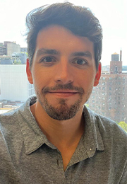
Juan Turati, Ph.D.
Role: Postdoctoral Fellow
Email: juan.turati@mssm.edu
Juan Turati, Ph.D., obtained his M.Sc. in biotechnology at the Argentine University of the Company, UADE in Buenos Aires, Argentina in 2015 and received his Ph.D. in neuroscience at the University of Buenos Aires, Argentina in 2021. His doctoral research focused on the role of metabotropic glutamate receptors on memory retention in Alzheimer disease (AD) and brain aging. From 2021-2022, Juan worked at mAbXience SAU, conducting pharmaceutical validation experiments involving monoclonal antibodies and cell lines. In 2023, Juan joined the Horng Lab as a postdoctoral fellow. His current research focuses on the role of astrocyte-immune cell crosstalk in controlling acute and chronic stages of CNS autoimmune disease.
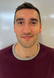
Josh Weiss, B.S.
Role: Research Associate
Email: josh.weiss@mssm.edu
Josh Weiss, B.S., graduated from the University of Maryland College Park in 2022 where he majored in Psychology. He is interested in the cellular and molecular mechanisms of neurological disorders, including multiple sclerosis, and intends to pursue an MD degree.

Jorge Villavicencio, BA
Role: Research Associate
Email: jorge.villavicencio@mssm.edu
Jorge Villavicencio received his BA from Rutgers University – New Brunswick with a major in Cell Biology and Neuroscience and a minor in Public Health. He previously worked on the maintenance and differentiation of hESCs and iPSCs in the Cohen Stem Cell Laboratory of the Rutgers Biomedical Engineering Department. There, he contributed to the development of differentiation protocols for myocardiocytes and neurons, and helped to refine a lentiviral transduction technique of VSV-Spike, which replaces the traditional glycoprotein with the SARS-CoV-2 Spike protein. Jorge plans to attend medical school after gaining more research experience in the Horng Laboratory during this gap year.
Alumini
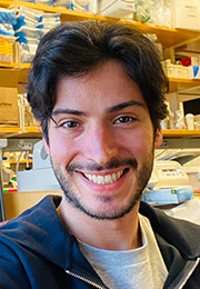
Mario Amatruda, Ph.D.
Role: Postdoctoral Fellow
Email: mario.amatruda@mssm.edu
Mario Amatruda, Ph.D., obtained his M.Sc. degree in biotechnology at the University Vita-Salute San Raffaele (Milan, Italy) in 2012. During his M.Sc., Mario investigated the role of neural progenitor cells in regulating the integrity of the blood-brain barrier in physiological conditions and after ischemic stroke. In 2017, Mario received his Ph.D. in neuroscience at the University College London (UK). His doctoral research was focused on the role of tissue hypoxia and energy deficit in multiple sclerosis (MS). During his PhD, Mario had additional training in drug screening at Keregen Therapeutics Ltd. (Stevenage, UK), where he examined the effects of Nrf2 activators for the treatment of MS. In 2017, Mario joined the Advanced Science Research Center at The City University of New York (New York, US). There, he studied the mechanisms contributing to the neurodegeneration in MS, and in particular the role played by specific signaling sphingolipids whose levels are altered in the brain of patients with MS. In 2020, Mario joined the Horng Lab as a postdoctoral fellow. His research focused on the role of astrocytic immune cell receptors in controlling acute and chronic patterns of CNS autoimmune disease. Mario now leads his own research team as a Senior Scientist at Biogen.
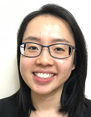
Viola Woo, BS
Role: Research Associate
Email: viola.woo@mssm.edu
Viola obtained her B.S. degree in biomedical sciences from SUNY Buffalo. She previously studied cocaine addiction and inflammatory bowel disease using mouse models and has also worked in drug development for autoimmune diseases. She is currently pursuing a degree in public health.

Joy Zhang, BS
Role: Research Associate
Email: joy.zhang@mssm.edu
Joy Zhang graduated from Duke University with a B.S. in Biology. At Duke, she studied dietary differences among the deer mouse populations of various California Channel Islands using stable isotope analysis. As an undergraduate, she also performed research on molecular mechanisms and translational approaches to retinal neovascularization in diabetic retinopathy at the Henry Ford Hospital. She plans to matriculate at medical school in the fall of 2019. She is currently pursuing her MD at the University of Virginia.
Positions
Postdoctoral Scientists
The Horng Laboratory of CNS Barriers and Neuroinflammation is searching for creative, hard-working Postdoctoral Scientists with a solid background in Neuroscience, Molecular Biology, Glial Biology, Mouse Genetics and/or Neuroimmunology. Experience in molecular biology, rodent handling, microscopy and basic biochemical techniques is required. Individuals with expertise in Astrocyte Biology, Immune Cell Differentiation, and/or RNA Sequencing are particularly encouraged to apply. Applicants should possess a Ph.D. and/or M.D. degree.
Please send a cover letter, curriculum vitae, and the names, phone numbers and e-mail addresses of three people who could provide letters of reference to sam.horng@mssm.edu.


