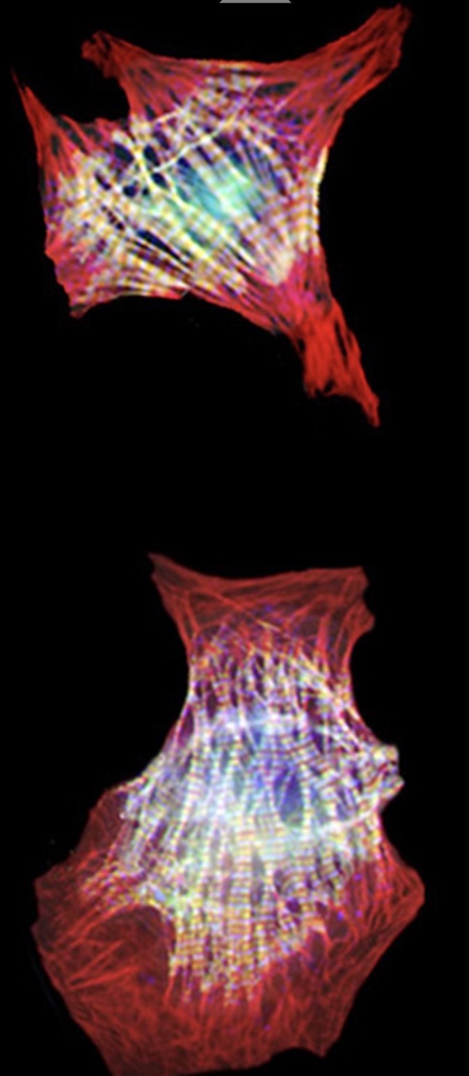
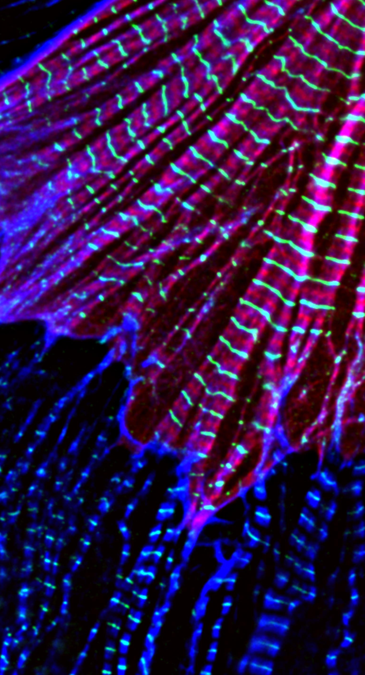
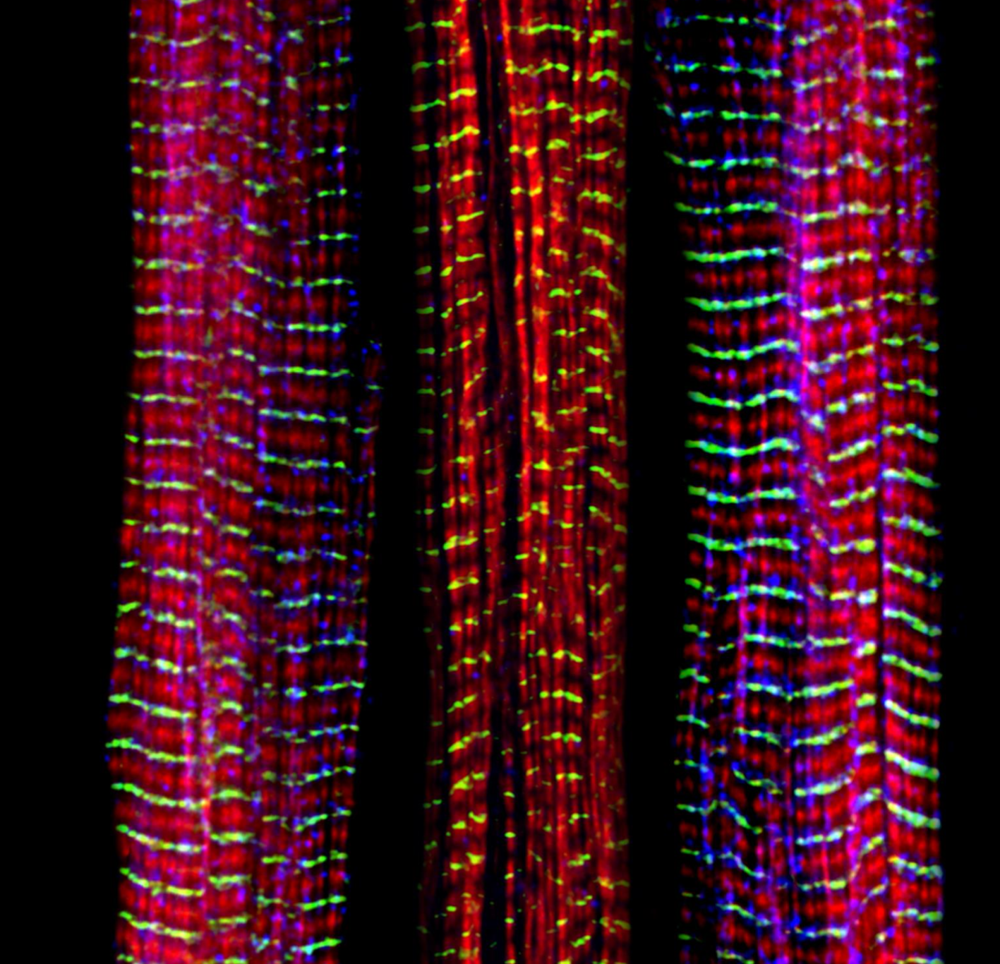
The research in this laboratory is focused on identifying the components and molecular mechanisms regulating actin architecture in cardiac and skeletal muscle during normal development and disease.
Control of actin filament lengths and dynamics is important for cell motility and architecture and is regulated in part by capping proteins that block elongation and depolymerization at both the fast-growing (barbed) and slow-growing (pointed) ends of the filaments.


Striated muscle is an ideal model system to test for the functional properties of various actin regulatory proteins due to the precise organization and polarity of cytoskeletal components within repeating sarcomeric units.
For example, the ~1 mm long actin filaments are easily resolved by light microscopy; thus we can make filaments longer and/or shorter and see the alterations by fluorescent microscopy in live myocytes.
Using this system we can combine advanced cell biological and biochemical approaches with direct tests of physiological function in live beating muscle cells.
The research objectives of the laboratory can be broadly summarized as follows:
1. Understanding the cellular mechanisms involved in the assembly, regulation and maintenance of contractile proteins in cardiac muscle in health and disease;
2. Deciphering the mechanisms critical for precisely specifying and maintaining the lengths of actin filaments. Actin is an indispensable structural element of cells and is the major component of heart muscle. Changes in actin, caused by genetic mutations, which have been identified in humans, are a frequent cause of several forms of cardiomyopathy. We are determining how genetic defects in this protein affect muscle force generation and muscle contraction, leading to sudden cardiac death.
3. Investigating how striated muscle cells maintain their shape, drive contraction, and generate mechanical force through the efficient integration of microfilament, microtubule and intermediate filament (IF) function.
4. Discovery of novel models of de novo cardiac muscle assembly, with special emphasis on differentiating murine embryonic stem (ES) cells to study the functional properties of specific cytoskeletal proteins (via “knock out” and “knock in” approaches) during all stages of heart muscle development.

