Research
Our lab is interested in understanding the mechanisms required for proper formation of the complex 4-chambered heart, and how errors in these processes cause congenital heart disease. The heart is composed of many different cell types, all of them essential for its proper function throughout the life of an organism. How the individual cell types are generated during development, and how they are selectively affected during disease remains a field with many open questions.
We use a wide combination of approaches to study these questions, including genetic mouse models, pluripotent stem cell differentiations and computational tools. Our overarching goals are to gain insight into cardiac lineage specification, morphogenesis and maturation to better understand and treat diseases of the heart.
We pursue our work in a vibrant and fun community of researchers interested in developmental, stem cell and cardiac biology, as well as the strong complementary expertise of the clinical community at Mount Sinai:
Dubois Laboratory
Nicole Dubois, Ph.D.
Associate Professor
Cell, Developmental and Regenerative Biology
Location
Lab: Hess 8-302
Office: Hess 8-112
Contact
Phone: 212-824-8546
Email: nicole.dubois@mssm.edu
Research Projects
Specification of Cardiac Lineages
Background and Impact: Congenital heart defects remain the most frequent birth defects in the human population, with 1% of newborns affected. The origin for many of these defects is thought to occur early during development, when the cardiac progenitor cells are specified. Identifying the key players and the mechanisms involved in early heart development will likely contribute to a better understanding of congenital heart defects, their characterization and diagnosis, and ultimately enhanced treatment opportunities.
Projects: We have identified a population of cells that give rise specifically to the ventricular chambers of the heart. These cells are labelled by expression of the transcription factor Foxa2 at gastrulation. We have further shown that FOXA2 also labels ventricular precursor cells during pluripotent stem cell differentiation. Lastly, we have used single cell RNA sequencing to comprehensively profile the cardiac progenitors and their differentiation trajectories in the early murine heart. In ongoing projects we expand on these studies to better understand 1) The heterogeneity of early cardiac progenitor populations, 2) Their spatio-temporal emergence, 3) The genes and pathways that are required for their specification, and 4) How different progenitor populations are affected when early development is perturbed, such as for example by high exposure to retinoic acid or other teratogens.
Approach and Technologies: We deploy the mouse embryo and lineage-tracing models, single cell sequencing technologies, CRISPR gene editing screens in pluripotent stem cell-derived atrial and ventricular cardiomyocytes, and more recently the use of complex organoid and bioprinted and engineered tissue models.
Mechanisms of de novo Sarcomerogenesis
Background and Impact: Sarcomeres are the building blocks of myofilaments, and exist in highly ordered patterns in cardiomyocytes. While the structure in mature cardiomyocytes has been studied extensively, how myofilaments are established at the very beginning, during de novo sarcomerogenesis, remains poorly understood. Identifying the detailed mechanisms of how cells established such intricate and highly ordered subcellular structures during development is a fascinating problem. Identifying the underlying concepts involved promises to reveal complex processes of cell biology and subcellular differentiation mechanisms, which may contribute to our understanding of sarcomere malformation, sarcomere malfunction, and sarcomere re-formation in regenerative models.
Projects: Here we are taking advantage of the fact that pluripotent stem cell differentiations closely recapitulate early heart development, including the process of sequential formation of sarcomeres. In a combined transcriptomic and proteomic screen we have identified processes at play across at all stages of sarcomere formation, including formation of Z bodies, formation of Z discs and maturation of myofilaments. In ongoing work we focus on 1) Understanding the role of the Z body as a biomolecular condensate, 2) Identifying candidates involved in local translation at the sarcomeres, 3) Identifying the subcellular structures and pathways involved in Z body formation, and 4) understanding how mutations in key sarcomere proteins impact de novo sarcomerogenesis.
Approach and Technologies: For this work we capitalize on the highly versatile PSC differentiation model, including PSC reporter lines, PSC gene editing, super resolution microscopy, FRAP, and live imaging, in combination with multiomic analysis such as proteomics, transcriptomics and eCLIP.
The Role of RNA Binding Proteins in the Heart
Background and Impact: The gene regulatory mechanisms driving early cardiac specification and differentiation have been studied extensively, and provided in depth insight into mechanisms of early heart development. Interestingly, the posttranscriptional regulation of these same processes is equally essential, yet much less studied, despite the fact that multiple RNA binding proteins have been identified in individuals with congenital heart defects. Identifying the key regulators involved in early heart development, and the mechanisms by which they function, promises to reveal additional regulatory concepts of early heart development, and relatedly, new insights into formation and mechanisms of congenital heart defects.
Projects: From a recent combined transcriptomics and proteomics screen we have identified multiple RNA binding proteins expressed during early heart development, including DDX3X. Mutations in DDX3X cause DDX3X syndrome, which manifests in neurodevelopmental defects and congenital heart defects in a subset of the patient population. To better understand how DDX3X contributes to congenital heart defects, ongoing work focuses on 1) Studying the consequence of Ddx3x loss-of-function during heart development, 2) The mechanism of action of DDX3X, and 3) The role of patient-specific mutations in DDX3X.
Approach and Technologies: To better understand the role of DDX3X in both mouse and human cardiac development we deploy classical mouse genetics and embryo studies, induced pluripotent stem cell biology and multiple technologies to assess RNA metabolism such as RNA-IP, eCLIP, Ribo-Seq and RNA imaging technologies.
Development and Disease of the Ventricular Conduction System
Background and Impact: Cardiac Purkinje Fibers cells are suspected to be a major contributor to ventricular arrhythmias, yet a detailed mechanistic understanding of how they do so, particularly in humans, remains largely elusive. Reasons for this knowledge gap include a paucity of animal models that specifically target the Purkinje Fiber cells (PFs), a lack of markers to isolate and study PFs and, importantly, the absence of human models with which to study the cellular and molecular mechanisms of PF physiology. As a consequence, clinical practice of ventricular arrhythmias currently does not provide strategies that address any cell-type specific (conduction versus working myocardium) involvement in disease. Classical animal models have provided important insights into the development of the PF system, via lineage-tracing and loss-of-function studies. However, defects in PF function, particularly those observed in the human population, are not readily recapitulated in such animal models. In recent years human pluripotent stem cell models have enabled the in vitro generation of atrial and ventricular cardiomyocytes, endocardium, epicardium and fibroblast cells, establishing platforms for disease modeling, cell therapy and drug discovery. Only few reports exist on generating Purkinje Fibers from pluripotent stem cells however, and none from human PSCs.
Project: Specifically, we study how the Purkinje Fiber cells are formed, both in vivo and in vitro, and how they distinguish themselves from working cardiomyocytes and from other conduction system cells such as pacemaker cells. We have found that transient induction of Notch signaling stably converts hPSC-derived cardiomyocytes (hPSC-CMs) to a Purkinje fiber phenotype. They adopt changes in morphology characteristic for PFs, and exhibit increased conduction and upstroke velocity, as well as a longer action potential duration compared to hPSC-CMs. Gene expression analysis further confirms a PF identity of Notch-induced cells as evidenced by up-regulation of classical PF markers and biological processes after conversion. Based on this new model system we now can ask important questions such as 1) How do Purkinje Fiber cells differ from working cardiomyocytes, 2) How to Purkinje Fibers interact with cardiomyocytes at the Purkinje fiber-myocyte junction?, 3) How do Purkinje Fiber cells behave in the context of disease, and 4) Can we predict Purkinje Fiber behavior based on computational models? In complementation to the in vitro studies we interrogate the Purkinje Fiber network in the mouse and ask the question of how these cells specify and mature over time.
Approach and Technologies: For this work we use an array of in vivo (mouse) and in vitro (hPSC) technologies, combined with electrophysiology analysis, single cell sequencing technologies, computational modeling and most recently, 3D bioprinting to generate Purkinje Fiber-myocyte mixed tissues.
Metabolism and Cardiac Maturation
Background and Impact: After specification and differentiation of the cardiac lineages, the heart undergoes a complex process of growth and maturation, that is not fully completed until several weeks after birth in the mouse, and years after birth in humans. One of the major changes that accompanies cardiac maturation is the change in metabolic activity. The fetal heart uses primarily glucose and lactate for fuel, while the postnatal heart relies mostly on fatty acids. What regulates this so called metabolic switch, and how metabolism more generally impacts the processes of cardiac growth and maturation is a fascinating area of investigation in our field. A better understanding of how metabolic activity is regulated, its interplay with cardiac growth and contractility, and the avenues by which it can be perturbed will impact the understanding of metabolic disorders, the establishment of better in vitro models and our understanding of metabolism in development and disease more broadly.
Project: We have recently shown that the metabolic switch that occurs in vivo can be recapitulated in vitro, in cardiomyocytes differentiated from pluripotent stem cells. Specifically, we found that the metabolic switch can be induced by activating PPARD, a pathway known to be involved in regulating metabolism in the adult heart. Activation of PPARD results in an increase in fatty acid oxidation, up-regulation of the fatty oxidation transcriptional machinery, in structural maturation of sarcomeres and in enhanced contractility of engineered tissues. Future work on this area I the lab includes the use of the enhanced maturation protocol to 1) Further study the mechanism underlying the activation of PPAR signaling in cardiomyocytes, and other cell types, 2) Study the genotype-phenotype correlations of lipid metabolism disorders, and 3) Deploy PPARD induced matured cardiomyocytes across various 3D cardiac tissues to test their impact on in vitro disease modeling and in vivo regenerative transplantation approaches.
Approach and Technologies: Here we deployed several standard cardiac biology assays, including imaging of sarcomere and mitochondrial structures, gene expression studies, measurement of cardiac contractility in engineered tissues and cellular metabolism such as seahorse and fatty acid flux assay. Additionally we generated iPSC lines with known patient mutations in metabolic enzymes and established 3D bioprinting assays.
Generating a Contractile Conduit for Single Ventricle Disease (Additional Ventures Cures Collaborative)
Background and Impact: Despite tremendous advancements in medicine in recent decades, the standard clinical approach for single ventricle disease has barely changed in the last 50 years. It consists of a series of open-heart surgeries over several years (Norwood procedure, Glenn operation, and Fontan procedure), which reconfigure the heart and its circulatory systems. While these surgeries are lifesaving, they do not fully restore normal cardiac physiology. The resulting non-pulsatile pulmonary circulation with passive flow leads to significant systemic complications, including in the lung, liver, kidneys and gastrointestinal tract, which typically culminates in multi-organ failure. Because of this, SVD patients have a shortened life span and a severely reduced quality of life overall.
Project: Our lab is one of 9 members of the Additional Ventures Cures Collaborative (AVCC), established in 2021 (https://www.additionalventures.org/initiatives/biomedical-research/cures-collaborative). The goal of the AVCC is to generate a contractile conduit from pluripotent stem cell-derived cardiomyocytes to be implanted in SVD patients in lieu of the non-contractile tube that is currently used. The role of our lab in the AVCC is to help generate highly functional and mature cells for the conduit, including cells generated via various maturation strategies and cells of the different lineages of the heart that are required to generated a maximally contractile and long-lasting conduit at human scale.
Approach and Technologies: The collective AVCC teams use a series of innovative and highly complementary approaches, which span the spectrum of basic heart development and stem cell technologies (our lab), cell production and scaling in bioreactors, electrophysiology expertise, state-of-the-art 3D bioprinting, computational modeling, small and large animal models and expertise from physicians-scientists treating SVD patients.
Publications
Shewale B, Ebrahim T, Samal A, Dubois NC. Molecular Regulation of Cardiomyocyte Maturation. Current Cardiology Reports. 2025 Jan 21;27(1):32. https://link.springer.com/article/10.1007/s11886-024-02189-1
Ebrahim TAM, Richter F, Gelb BD, Dubois NC. The Heart from Every Angle Recent Approaches To Modeling Congenital Heart Disease. Current Treatment Options in Cardiovascular Medicine. 2025 Oct 16;27(1):69. https://link.springer.com/article/10.1007/s11936-025-01123-0
Gonzalez DM, Schrode N, Ebrahim TAM, Broguiere N, Rossi G, Drakhlis L, Zweigerdt R, Lutolf MP, Beaumont KG, Sebra R, Dubois NC. Dissecting mechanisms of chamber-specific cardiac differentiation and its perturbation following retinoic acid exposure. Development. 2022 Jul 1;149(13):dev200557. https://journals.biologists.com/dev/article/149/13/dev200557/275942/Dissecting-mechanisms-of-chamber-specific-cardiac
Wickramasinghe NM, Sachs D, Shewale B, Gonzalez DM, Dhanan-Krishnan P, Torre D, LaMarca E, Raimo S, Dariolli R, Serasinghe MN, Mayourian J, Sebra R, Beaumont K, Iyengar S, French DL, Hansen A, Eschenhagen T, Chipuk JE, Sobie EA, Jacobs A, Akbarian S, Ischiropoulos H, Ma’ayan A, Houten SM, Costa K, Dubois NC. PPARdelta activation induces metabolic and contractile maturation of human pluripotent stem cell-derived cardiomyocytes. Cell Stem Cell. 2022 Apr 7;29(4):559-576.e7. https://www.cell.com/cell-stem-cell/fulltext/S1934-5909(22)00097-2?keyword=diabetes
Shewale B, Dubois N. Of form and function: Early cardiac morphogenesis across classical and emerging model systems. Semin Cell Dev Biol. 2021 Oct;118:107-118. https://www.sciencedirect.com/science/article/pii/S1084952121000987
Bardot ES, Dubois NC. A Watershed Finding for Heart Regeneration. Cell. 2019 Feb 21;176(5):947-949. https://www.cell.com/cell/fulltext/S0092-8674(19)30116-3
Bardot E, Tzavaras N, Benson DL, Dubois NC. Quantitative Whole-mount Immunofluorescence Analysis of Cardiac Progenitor Populations in Mouse Embryos. J Vis Exp. 2017 Oct 12;(128):10.3791/56446. https://app.jove.com/t/56446/quantitative-whole-mount-immunofluorescence-analysis-cardiac
Bardot E, Calderon D, Santoriello F, Han S, Cheung K, Jadhav B, Burtscher I, Artap S, Jain R, Epstein J, Lickert H, Gouon-Evans V, Sharp AJ, Dubois NC. Foxa2 identifies a cardiac progenitor population with ventricular differentiation potential. Nat Commun. 2017 Feb 14;8:14428. https://www.nature.com/articles/ncomms14428
Wyatt CM, Dubois N. In vitro generation of renal tubular epithelial cells from fibroblasts: implications for precision and regenerative medicine in nephrology. Kidney Int. 2017 Feb;91(2):265-267. https://www.sciencedirect.com/science/article/pii/S0085253816306913
Calderon D, Bardot E, Dubois N. Probing early heart development to instruct stem cell differentiation strategies. Dev Dyn. 2016 Dec;245(12):1130-1144. https://anatomypubs.onlinelibrary.wiley.com/doi/full/10.1002/dvdy.24441
Dubois NC, Craft AM, Sharma P, Elliott DA, Stanley EG, Elefanty AG,Gramolini A, Keller G. SIRPA is a specific cell-surface marker for isolating cardiomyocytes derived from human pluripotent stem cells. Nat Biotechnol. 2011 Oct 23;29(11):1011-8. https://www.nature.com/articles/nbt.2005
Dubois NC, Adolphe C, Ehninger A, Wang RA, Robertson EJ, Trumpp A. Placental rescue reveals a sole requirement for c-Myc in embryonic erythroblast survival and hematopoietic stem cell function. Development. 2008 Aug;135(14):2455-65. https://journals.biologists.com/dev/article/135/14/2455/43595/Placental-rescue-reveals-a-sole-requirement-for-c
Dubois NC, Hofmann D, Kaloulis K, Bishop JM, Trumpp A. Nestin-Cre transgenic mouse line Nes-Cre1 mediates highly efficient Cre/loxP mediated recombination in the nervous system, kidney, and somite-derived tissues. Genesis. 2006 Aug;44(8):355-60. https://onlinelibrary.wiley.com/doi/abs/10.1002/dvg.20226
Meet the Team
Nicole received her BA in cell biology from the Swiss Federal Institute of Technology (ETHZ) in Zurich, Switzerland, in 2003. She then moved to the lab of Dr. Andreas Trumpp at the Swiss Cancer Institute (ISREC) in Lausanne, where she studied the role of c-Myc, an oncogene, during development of the hematopoietic system, and where she obtained her PhD in Cell and Developmental Biology, from the University of Lausanne, Switzerland, in 2007. From 2007-2013 she worked in the lab of Dr. Gordon Keller at the McEwen Institute for Regenerative Medicine and the University of Toronto Health Network in Toronto to study early cardiac specification and differentiation mechanisms in pluripotent stem cell models. In 2013 Nicole started her own lab at the Icahn School of Medicine at Mount Sinai. Lab research continues to investigate the various mechanisms underlying early heart development and congenital heart defects, and Nicole enjoys sharing her passion for research and discovery, for collaboration, mentoring and making work a fun and impactful place with all of her lab.
Arushi received her BS in molecular and cellular biology from Johns Hopkins University. At Hopkins she worked in the lab of Dr. Katherine Wilson studying the interaction between nuclear envelope proteins and the nuclear lamina network in the context of chronic inflammation. Following her undergraduate studies she joined the PhD program at Mount Sinai and joined the Dubois Lab in 2024.
At Sinai she is involved in MiNDs, the PREP Mentorship program, and is a PhD mentor. Outside of lab she loves trying new restaurants in the city, running in Central park, and reading.
Alex received her B.S. in biology from Brown University. At Brown, she completed her honors thesis work in the lab of Dr. Elena Oancea studying signaling pathways in melanocytes. She then joined Monte Rosa Therapeutics, a small biotechnology company developing ‘molecular glue degrader’ therapeutics, as a research associate studying targeted protein degradation across various autoimmune and oncological applications.
Alex joined the Dubois Lab in 2025. She is interested in studying mechanisms of local translation at the cardiac sarcomere, particularly through the lens of RNA binding proteins. She enjoys scientific communication and has participated in coordinating outreach events through Mentoring in Neuroscience Discovery at Sinai (MiNDS) and Save Our Science. In her free time, Alex enjoys climbing, ultimate frisbee, and exploring Central Park.

Emily received her B.S. in Biomedical Engineering, summa cum laude, from Bucknell University. During her undergraduate career, she conducted research in the labs of Dr. Brandon Vogel and Dr. Olivia (Ngo) Boerman, designing novel hydrogel formulations for applications in synthetic extracellular matrices and drug delivery. She also gained industry experience at AbbVie Pharmaceuticals, where she worked in drug metabolism and contributed to the development of iPSC-derived models of the corneal epithelium.
Prior to joining Mount Sinai as a PhD student, Emily worked as an Associate Researcher in the Department of Neuroscience in Dr. Joel Blanchard’s lab. In this role, she investigated how APOE genotype influences phosphorylated tau accumulation in glial cells and contributes to Alzheimer’s disease risk and resilience using the iPSC-derived human brain tissue-like miBrain model system.
Emily joined the Dubois Lab in 2026. She is interested in understanding how interactions within the cardiac microenvironment are established and modulated across development and aging.
At Mount Sinai, she is a leader of the Get Empowered in Medicine and Science (GEMS) Committee and a member of the Consulting Club, Science Career Club, and Mentoring in Neuroscience Discovery at Sinai (MiNDS). In her free time, Emily enjoys running, spending time with friends and family, and rating NYC restaurants on the Beli app.
Alumni
Bhavana Shewale
Tasneem A. M. Ebrahim

Samudyatha Prasad
Ivianis Nieves Carril
Evan Bardot
Damelys Calderon

Priyanka Dhanan
Anna Popplestone
Nadeera Wickramasinghe
David Gonzalez
Frank Santoriello
Zeynep Cakmak
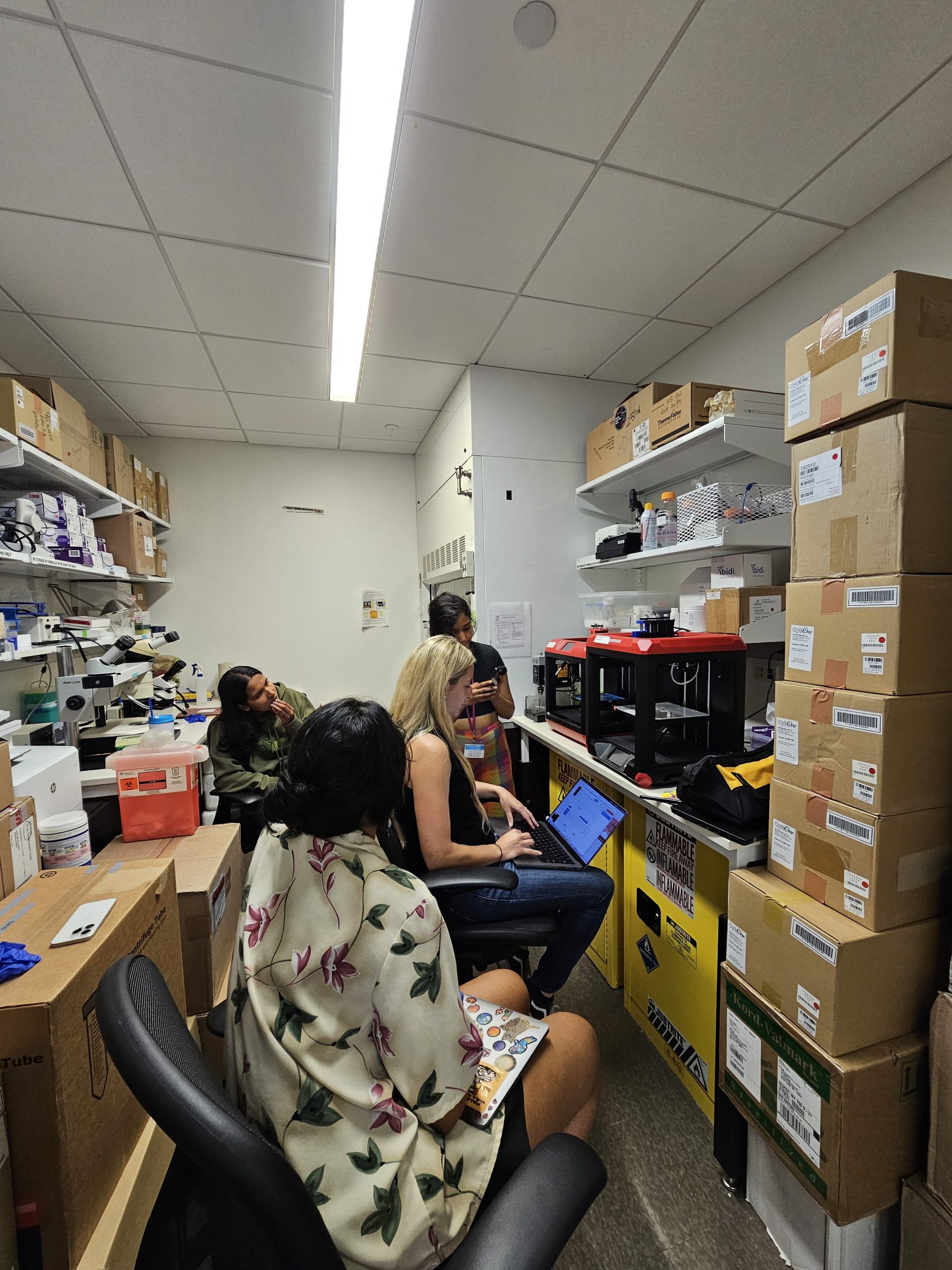
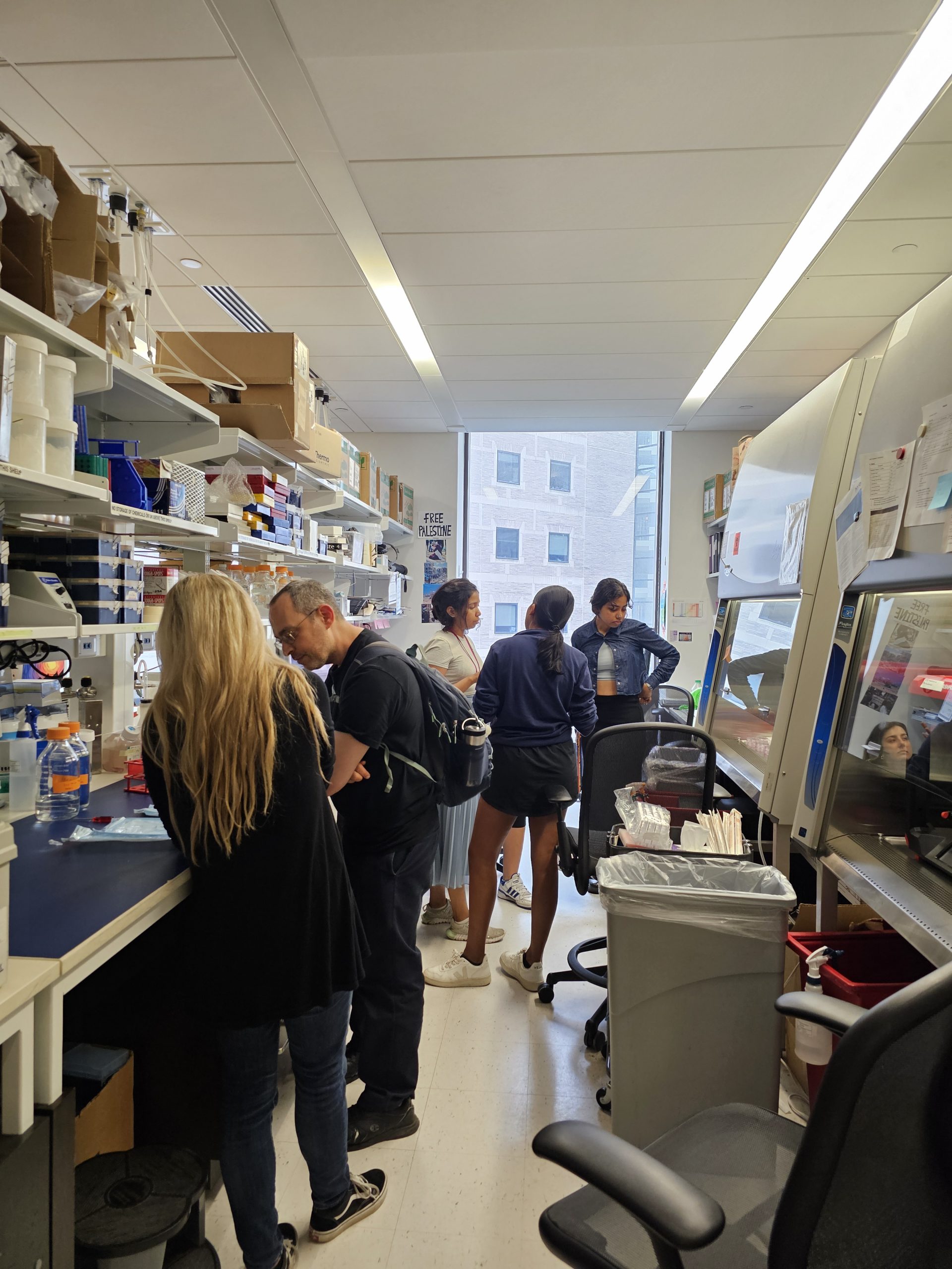
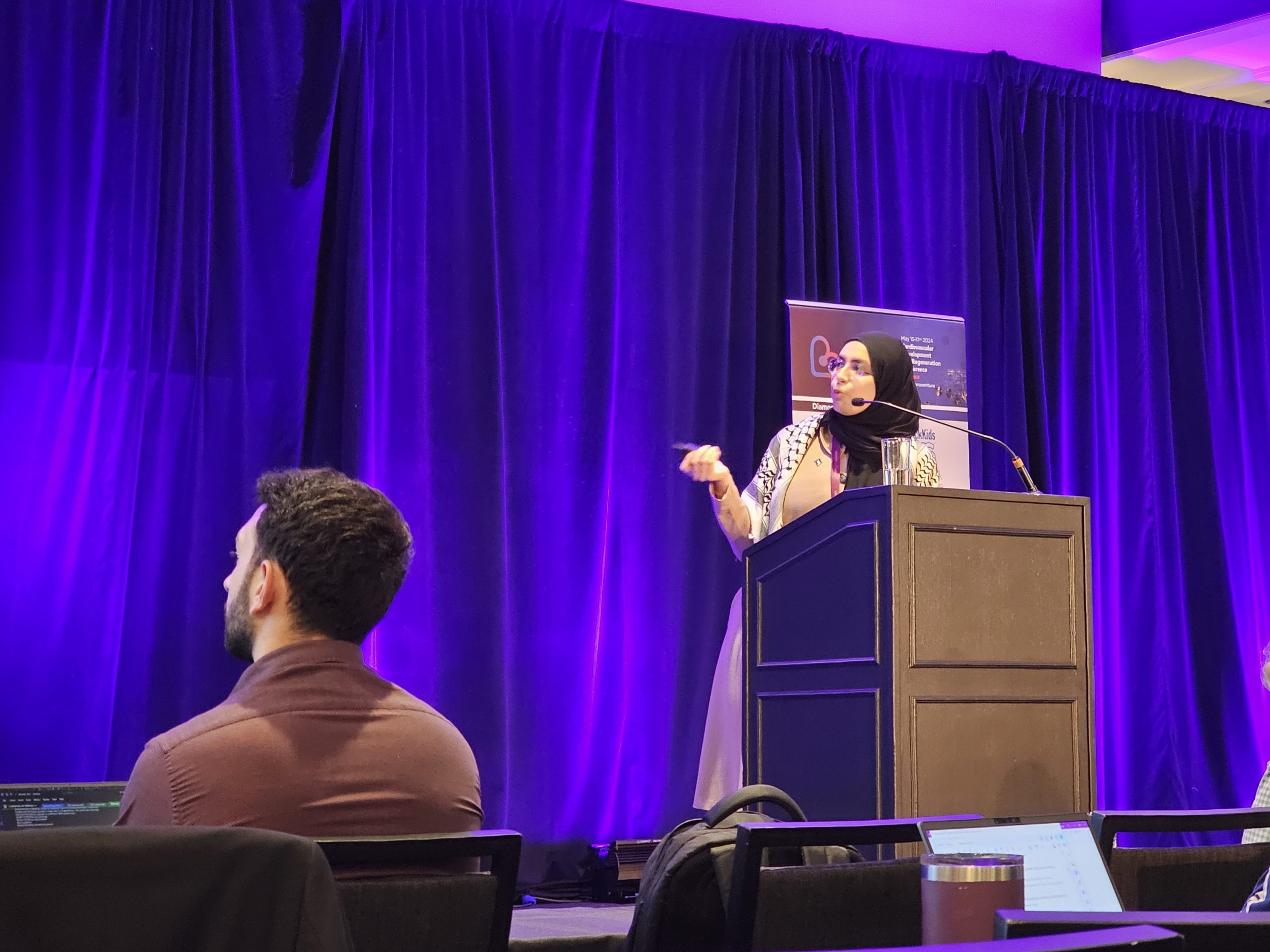
Gallery and News
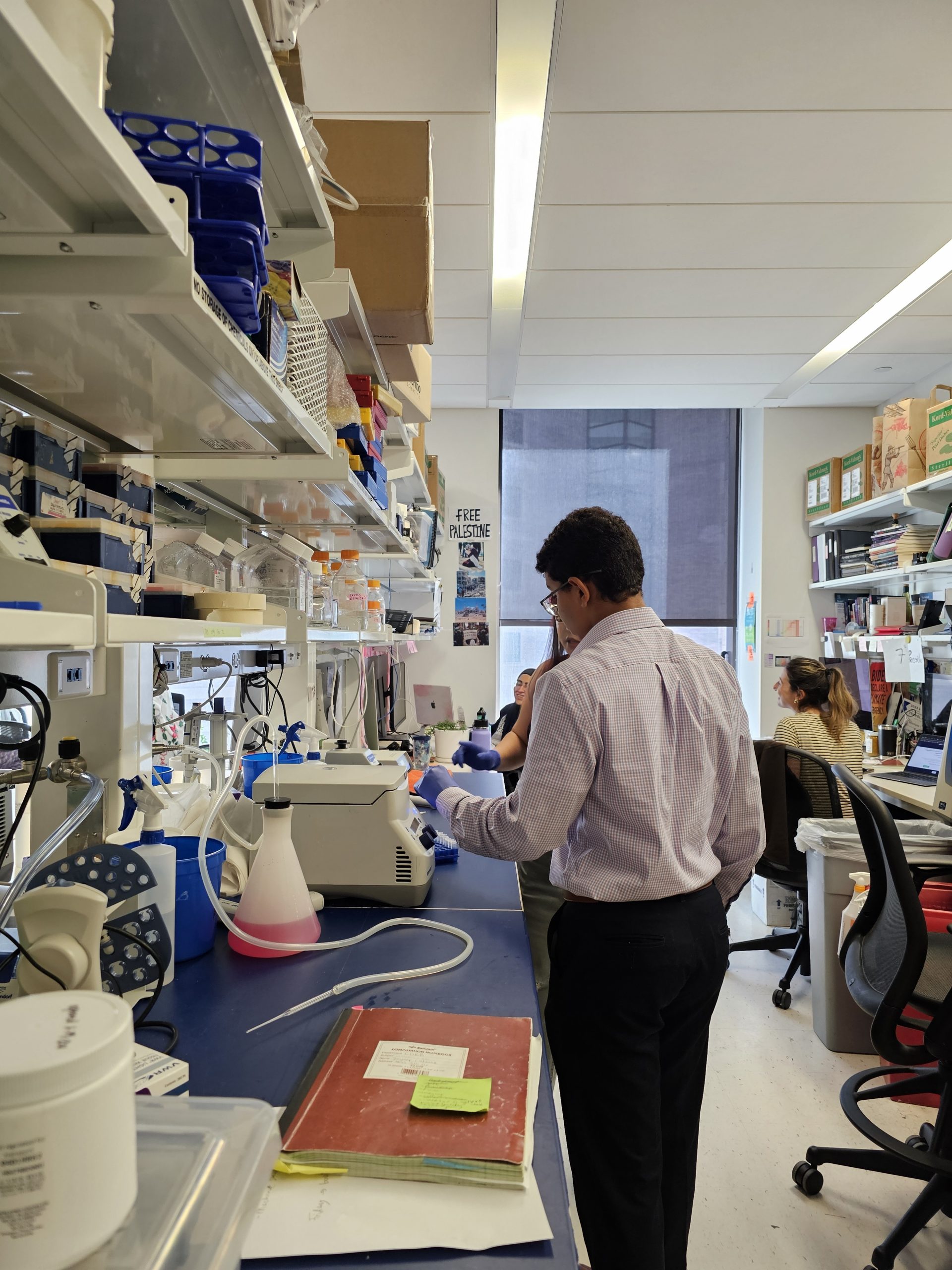
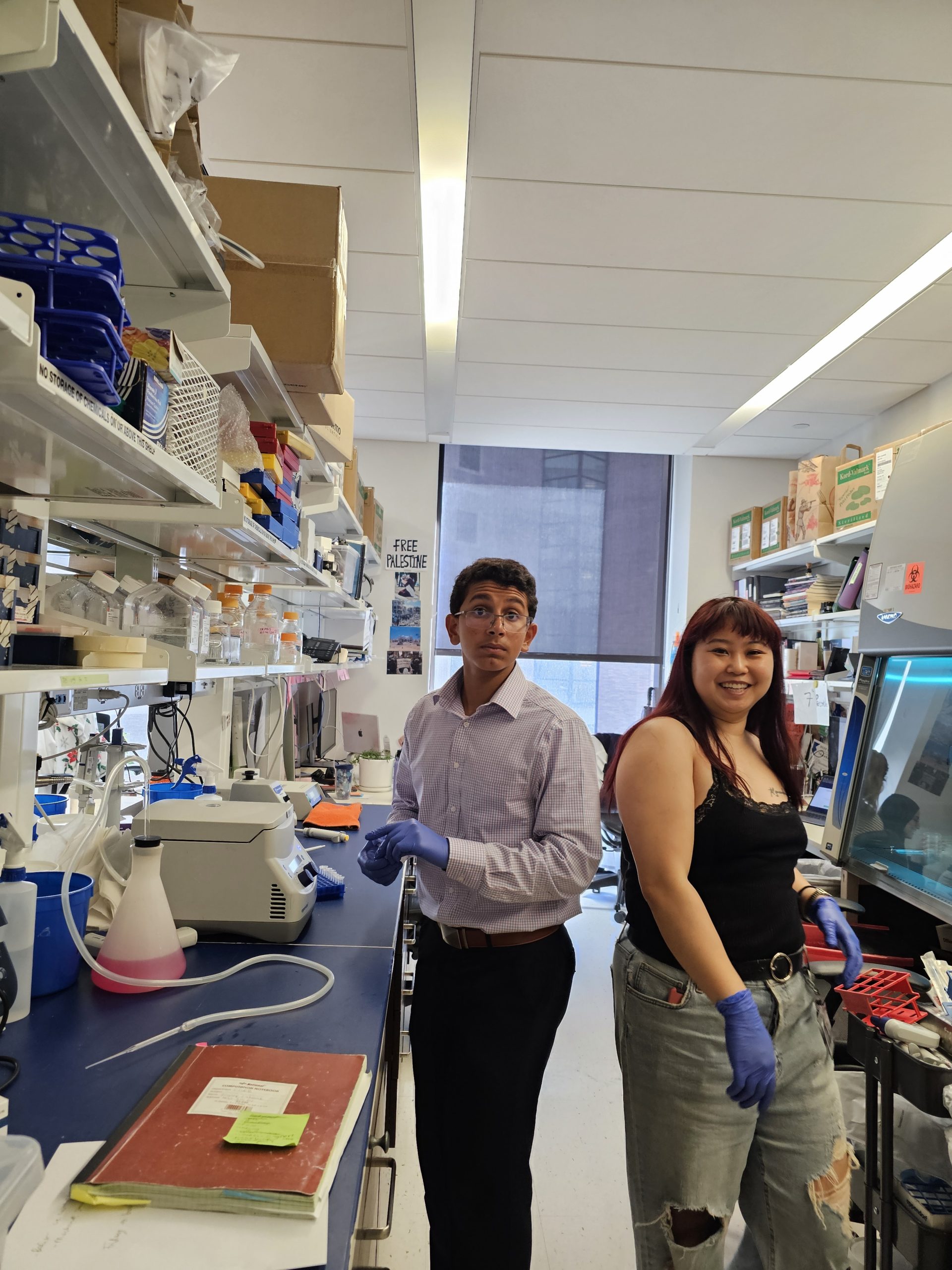
Jeremy joins the lab for the summer
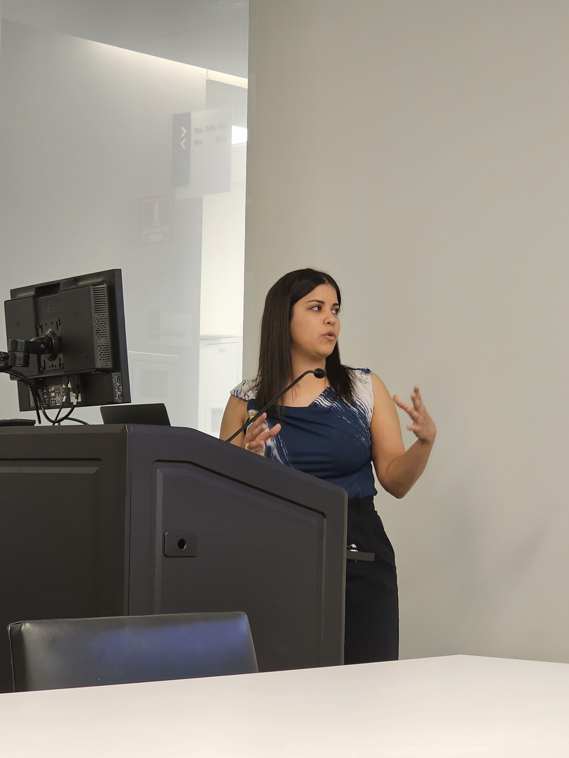
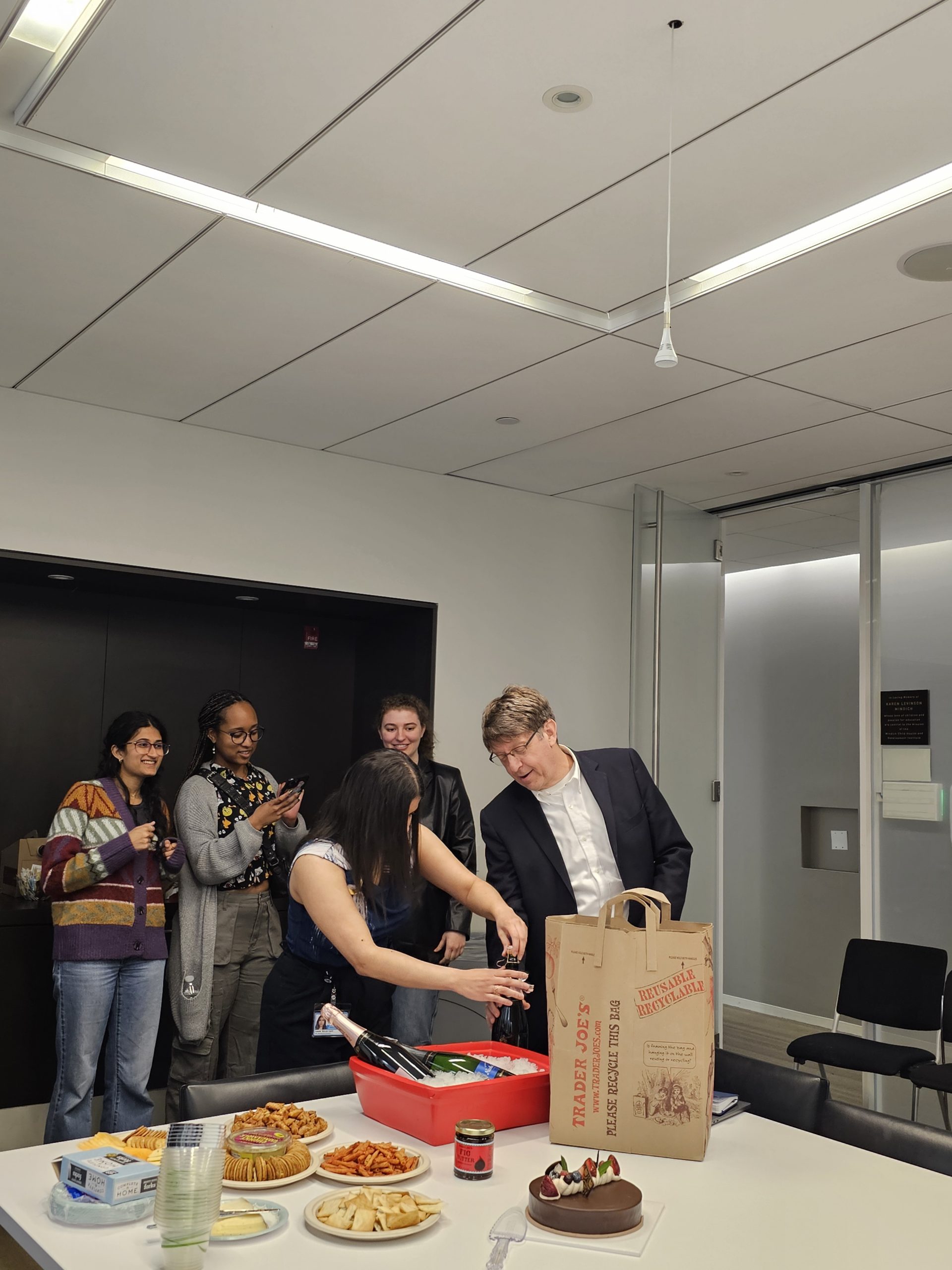
Ivianis’s defense

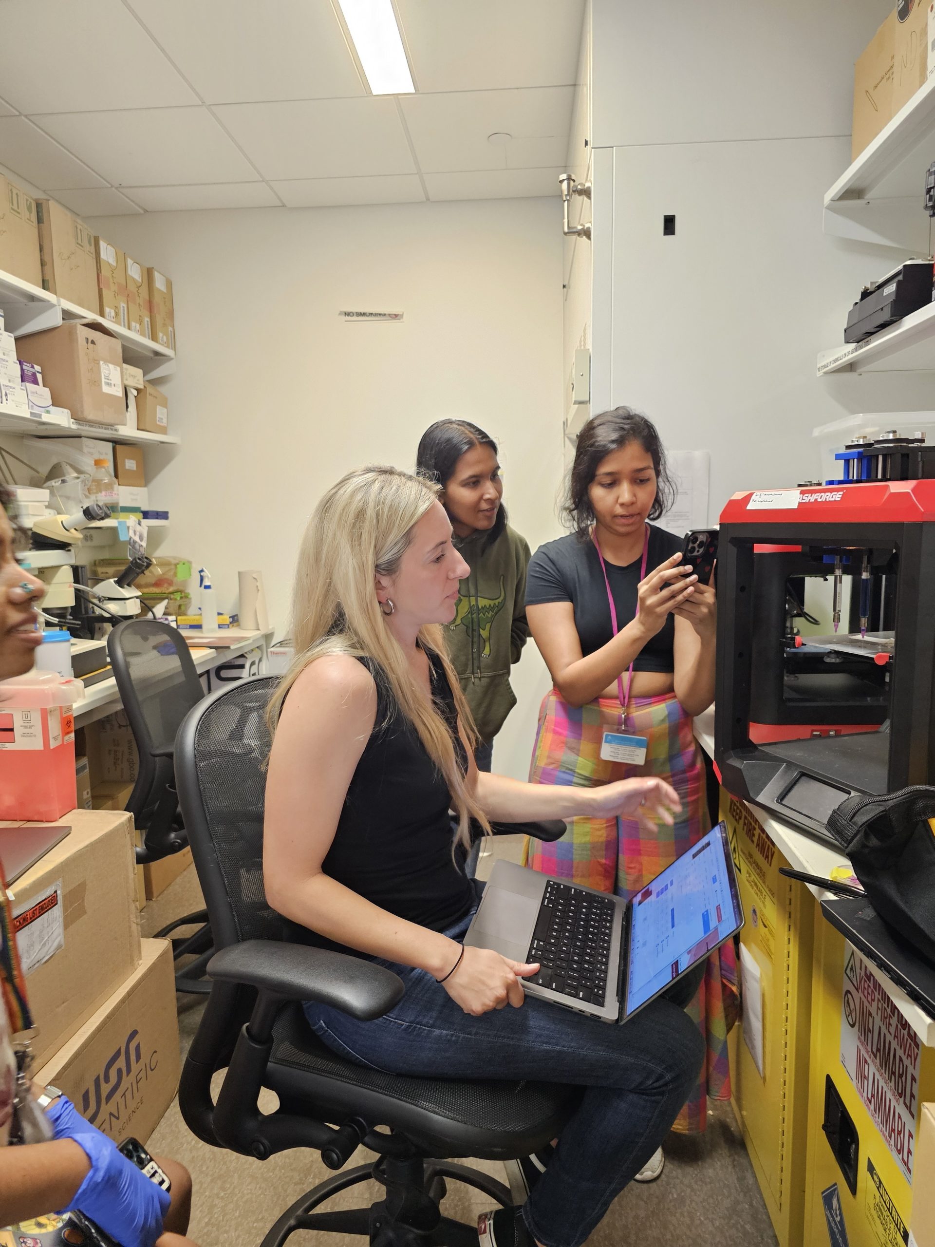
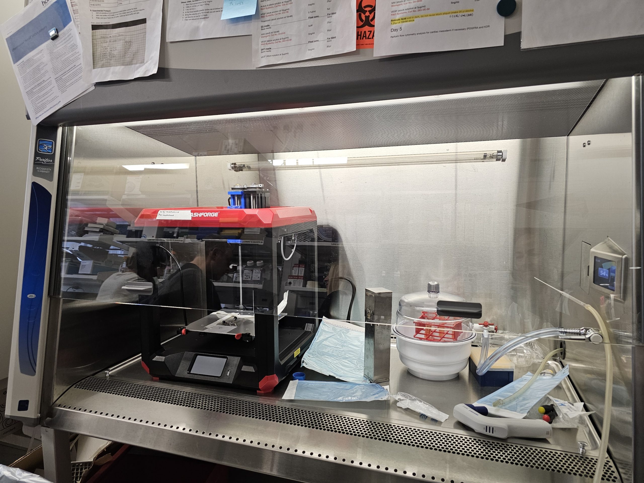
Jaci’s visit to set up 3D bioprinting
Weinstein 2024
Other Things we Care About
Education, at Sinai and Beyond
Fellowships and Funding Opportunities
Meetings and Conferences
Living in New York City
Dubois Laboratory
Location
Lab: Hess 8-302
Office: Hess 8-112
Contact
Phone: 212-824-8546
Email: nicole.dubois@mssm.edu