Research
The Beck lab uses advanced MRI techniques and ultra-high field MRI to understand how brain and spinal cord lesions form, grow, and repair in multiple sclerosis and other autoimmune disorders that affect the central nervous system. We couple advanced MRI techniques with analysis of CSF and tissue specimens to understand the mechanisms underlying lesion formation and repair. With this work, we hope to improve MS diagnostic and prognostic accuracy and develop new, more targeted treatments for people with MS.
How do lesions in the cortex form and repair in the cortex in multiple sclerosis?
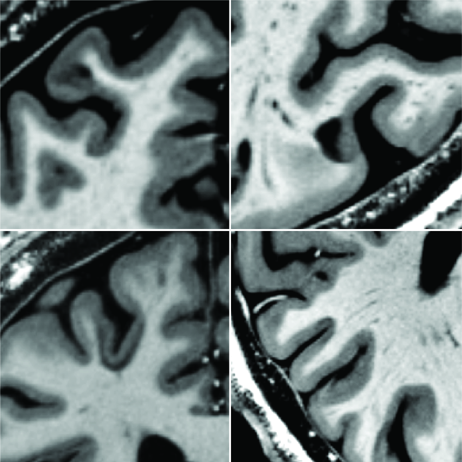
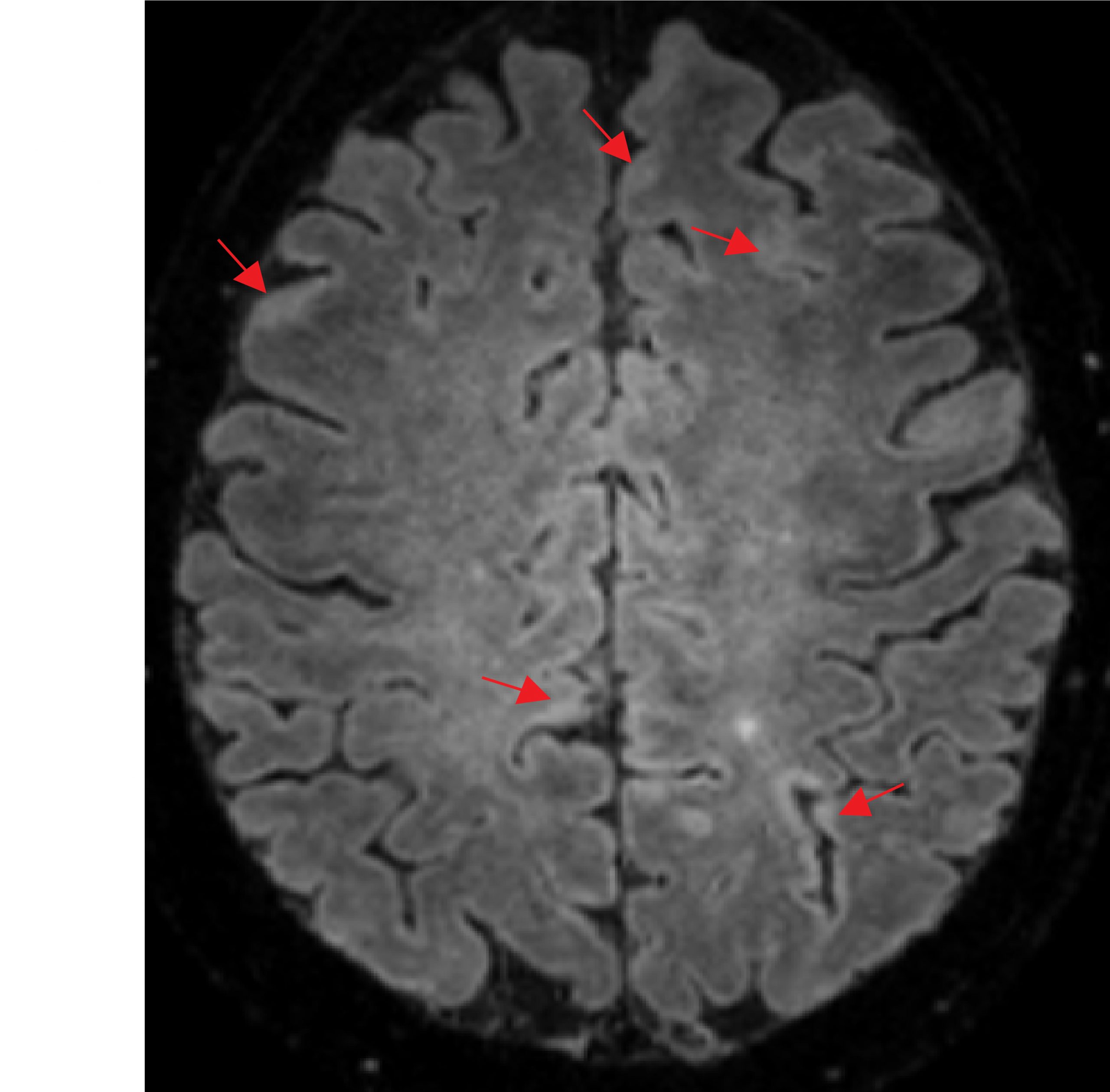
Although traditionally thought of as a disease of the white matter, lesions in the cerebral cortex are common and can be extensive in MS. Cortical lesions, however, are hard to see using standard MRI techniques and so we have developed multiple, advanced techniques using ultra-high field 7 tesla (7T) MRI to see these lesions (Beck et al, AJNR Am J Neuroradiol 2018, Beck et al, Invest Radiol 2021). With these techniques, we and others have demonstrated that cortical lesions are associated with disability and progressive MS (Beck et al, Mult Scler, 2022). In people with longstanding MS, we have found that cortical lesion formation is infrequent, but that cortical lesion burden is associated with worsening disability over time (Beck et al, Brain Commun, 2024). Our ongoing work seeks to understand when and in whom cortical lesions develop, how lesion formation and repair differ between the cortex and other parts of the central nervous system, and how cortical lesions are related to more diffuse MS pathology, including neurodegeneration and brain atrophy.


What are the mechanisms underlying cortical lesions in multiple sclerosis?
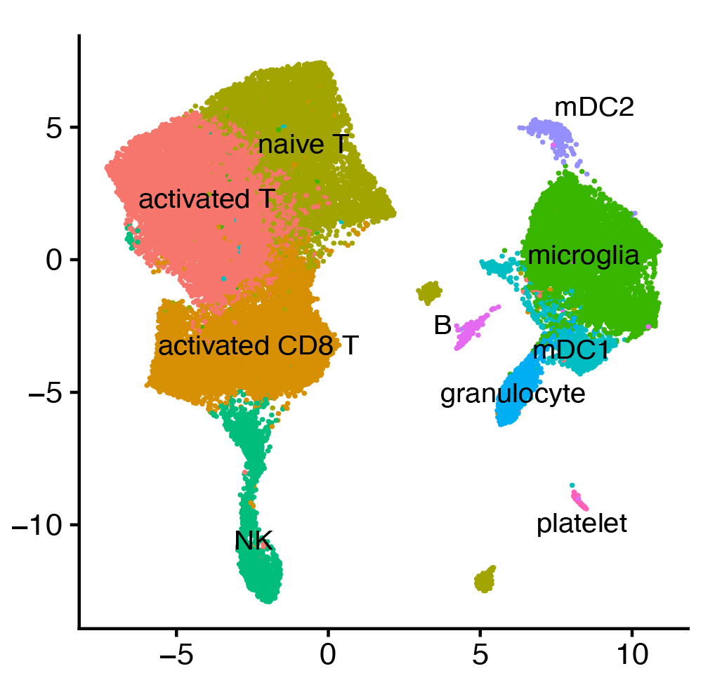
Cortical, in particular subpial cortical, lesions may arise due to inflammation in the overlying meninges, in contrast to lesions in the cerebral white matter. By studying the immune landscape of the cerebrospinal fluid in people with high vs low cortical lesion burden, using cutting edge transcriptomic techniques, we hope to understand what immune cells and pathways are implicated in cortical lesion formation. With this information, we may be better able to prevent these lesions from forming or promote their repair.
Development of new imaging and image analysis techniques
Although we have made great progress in developing imaging techniques for visualizing and characterizing MS lesions, we continue to work to improve on these techniques and develop new techniques, including a T2* weighted method that allows much more sensitive detection of cortical lesions at 3T (Beck et al, Invest Radiol 2021). We are working to improve and validate this technique, which will allow much larger scale clinical research on cortical lesions, and ultimately may facilitate the inclusion of cortical lesions in clinical trials and in routine clinical care. We are also working on image analysis techniques that will make cortical and white matter lesion identification easier, faster, and more accurate (La Rosa et al, ISMRM 2020; La Rosa et al, NMR Biomed 2022, Dereskewicz et al, J Neuroimaging 2025).
We have also developed a technique for estimating brain age directly from T1 weighted images (La Rosa et al, Imaging Neurosci 2025). We find that brain age is older than chronological age in people with MS and are studying the factors that influence brain age and how accelerated brain aging impacts disability in MS.

Translation of advanced MRI techniques to other autoimmune diseases that affect the central nervous system
MRI has been an incredibly powerful tool for understanding the pathophysiology of MS and developing treatments. We plan to translate some of the imaging techniques used in MS to other autoimmune and infectious disorders that affect the central nervous system in order to better understand the similarities and differences between MS and other autoimmune CNS disease as well as to improve diagnosis and care of these disorders.
